|
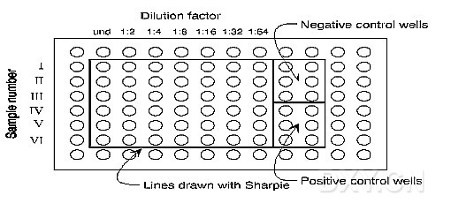
A.Sensitize a 96-well microtiter plate with purified antigen. 1.Prepare a solution of the purified antigen of interest in phosphate buffer (see recipe below) such that a concentration of approximately 10 µg/ml is achieved. Pipette 100 µl of this solution to all wells of the plate with the exception of those in the outermost rows and columns (these areas of the plate are prone to evaporation). 2.Cover the plate with Parafilm and incubate for 2 hours at 37°C, followed by an overnight incubation at 4°C. Cover the plate tightly to avoid evaporation. Store at 4°C. The plate(s) will remain usable for about one month. B.Block the sensitized plate. 1.When ready to use, discard the antigen solution contained in the wells of the sensitized plate. Pat the inverted plate sharply several times against a pad of paper towels to insure complete removal of the solution. 2.Wash the wells three times with 1x PBS/0.05%-Tween 20. Pat to dry completely after the final wash, as above. 3.Pipette 100 µl of 5% skim milk/PBS/0.005% thimerasol (added to retard bacterial growth) to each well. Incubate the plate, covered with Parafilm, at 37°C for 2 hours to overnight. It is convenient during this incubation step to begin preparation of the samples to be tested, outlined below. 4.Wash the wells three times with PBS/Tween, dry as before after the last wash. C.Prepare the samples to be tested. 1.Using a Sharpie or other suitable permenant marker, mark a second, unsensitized microtiter plate to identify the intended location of all samples to be tested, and the location of the positive and negative controls (the positive control in this assay is a purified sample of the antigen of interest; the same antigen with which the previous plate has been sensitized. The negative control is PBS). Do not use the outermost wells of the plate, as these have not been sensitized in the previous plate. See diagram. 2.Pipette 60 µl of 5% skim milk/PBS/Tween to all experimental wells except the first column of these wells, which will contain undiluted aliquots of the samples to be tested. 3.Add 60 µl of the samples to be tested to both the first (empty) and second wells of the experimental area of the plate. Mix the 1:2 dilution (second well) by pipetting up and down several times. 4.Using a fresh pipette tip, transfer 60 ul from well 2 (1:2 dilution) to well 3, creating a 1:4 dilution in this well. Mix by pipetting several times. Serially dilute the samples in this manner until all desired dilutions are achieved. It is critically important to use a fresh pipette tip for each transfer. 5.Place 60 µl of PBS in the negative control wells and 60 µl of the purified antigen sample in the positive control wells. 6.Dilute antisera to the antigen of interest appropriately in 5% skim milk/PBS/Tween (i.e. 1:1000, optimal dilution may vary depending on antigen-antisera. Final dilution under experimental conditions will be doubled) and pipette 60 µl of the diluted antisera to all wells (experimental and control). 7.Incubate the plate, covered with Parafilm to minimize evaporation, at 37°C for 2 hours to overnight. D.Testing the samples. 1.Retrieve the sensitized and blocked microtiter plate prepared in sections A & B, above. Duplicate exactly the pattern drawn on the non-sensitized plate on this plate. 2.Transfer 100 µl of the antisera-antigen solutions from the non-sensitized plate to their corresponding wells on the sensitized, blocked plate. Incubate this plate, covered, at 37°C for 1.5 hours. 3.Discard the solutions from the wells of the plate after incubation. Wash all wells three times with PBS/Tween as before. Dry after last wash. 4.Add to each well 100 µl of conjugate antibody (i.e. goat antirabbit alkaline phosphatase, if antisera was raised in rabbits) diluted appropriately in skim milk/PBS/Tween (i.e. 1:2000). Incubate the covered plate at 37°C for 1.5 hours. 5.Discard the conjugate antibody solution from the wells. Wash three times with PBS/Tween as before. 6.Add to each well 100 µl of substrate solution appropriate for the conjugate antibody used. For alkaline phosphatase conjugate, use p-nitrophenyl phosphate disodium (Sigma 104) dissolved in substrate buffer (795 mg Na2CO3, 1.456 g NaHCO3, 100 mg NaN3, 50 mg MgCl2 in a final volume of 500 ml sdH2O). 7.Read absorbence values at appropriate time intervals (usually every 15 minutes, a shorter interval is required if the color reaction is quicker; watch color development). Phosphate Buffer Skim Milk Solutions
Solution 1 Solution 2 50.0 g skim milk powder
Na2HPO4 2.84 g NaH2PO4 2.76 g 100.0 ml 10x PBS
dH2O to 100 ml dH2O to 100 ml 1.0 ml 5% thimerosal
sterile dH2O to final volume of 1L
Combine 6.1 ml of solution 1 and
3.9 ml of solution 2. Add 40 ml dH2O. Divide solution into 500 ml aliquots.
To one aliquot, add 250 µl Tween-20.
Pasturize at 65°C for 30 minutes.
10x Phosphate Buffered Saline (PBS) NaCl 80.0 g KCl 2.0 g Na2HPO4 14.4 g KH2PO4 2.4 g Dissolve reagents in 800 ml dH20. Adjust pH to 7.4 with HCl. Autoclave to sterilize. Dilute to 1x before use. Add 250 µl Tween to 500 ml 1x PBS for the PBS/Tween wash solution. |
→如果您认为本词条还有待完善,请 编辑词条
词条内容仅供参考,如果您需要解决具体问题
(尤其在法律、医学等领域),建议您咨询相关领域专业人士。
0
收藏到: