|
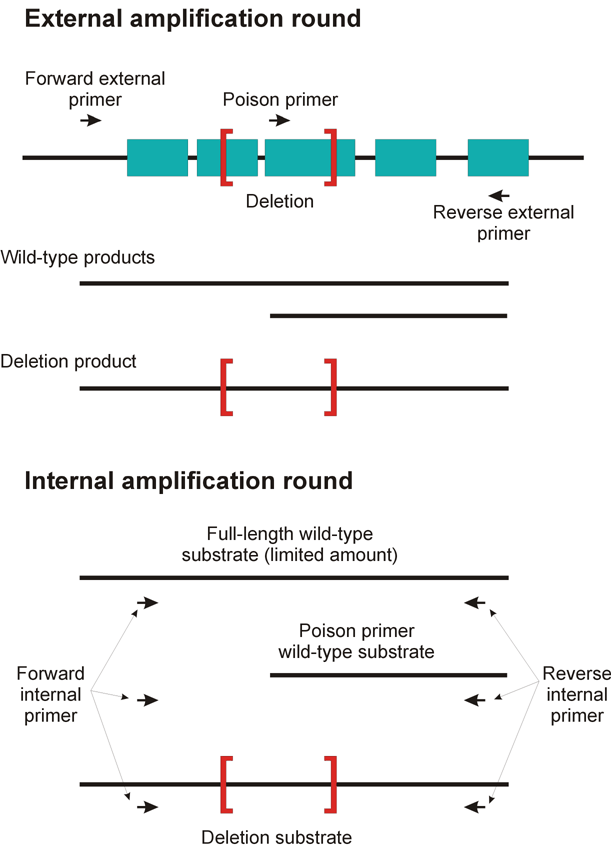
Introduction PCR protocols to detect rare deletions in DNA from complex populations of mutagenized worms typically depend on a large size differential between the wild-type product and the deletion product. These protocols are used with libraries in which deletion representation is about 1 chromosome in 20,000. Recent work has shown that a significant proportion of deletions induced by trimethyl psoralen (TMP) treatment followed by UV irradiation are small, on the order of 50 to 600 base pairs (Gilchrist et al., in preparation). Detecting these deletions via PCR presents a problem because deletion amplicons are not favored over wild-type products if the amplicons are similar in size. To circumvent this problem we use a "poison primer" technique for nested PCR based on a concept proposed by Gary Moulder and a protocol developed by Mark Edgley (Edgley et al., Nucleic Acids Res. 30(12):e52, Jun 15 2002). This method works because the poison allows the formation of deletion products but titers out full-sized products. The following material describes the method and shows some of our test results. How It Works In the first round of PCR, we include a third functional PCR primer that falls between the two external primers (Figure 1). Amplification from the wild-type template leads to the production of two fragments, one full-length and one relatively short. In practice, the shorter fragment is produced much more efficiently than the longer. Amplification from a mutant template, in which the site for the third internal primer is deleted, leads to the production of a single mutant fragment from the normal external primers. In the second round of PCR, we use two primers placed just inside the external first round primers. The shorter wild-type band from the first round cannot serve as a template for the second round PCR because it does not include one of the second round primer sites. The longer wild-type fragment can serve as a template, but because its production was limited by competition in the first round, its production in the second round is limited correspondingly. The lower level of effective wild type gives the deletion fragment an advantage.
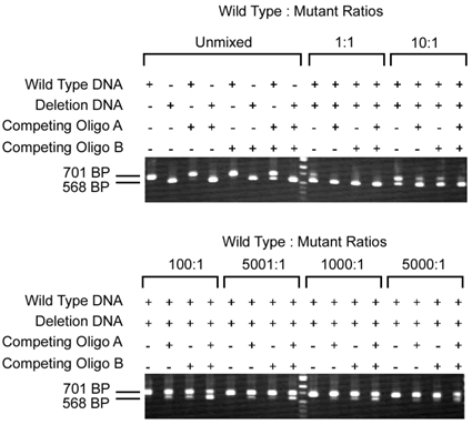
Figure 1. The poison primer technique. Introduction of a competing "poison" primer in the first round external PCR interfered with the production of a full length product. If the poison primer binding site is removed by a deletion, a full length PCR product can be enriched, allowing second round amplification of the deleted PCR product. Increased Sensitivity of Detection in DNA Mixing PCR Experiments We tested the poison primer method on a 133 bp deletion in the gene dim-1, using two different internal poison primers (A and B) that lie within the deletion interval (Figure 2). One or both were included in the external round of nested PCR, with different mixtures of wild type and deletion mutant DNAs used as the beginning template at the indicated ratios.With no poison primer, both the 701 bp wild type PCR fragment and 568 bp deletion fragment were observed in reactions with WT:Mutant ratios up to 10:1, but the ability to detect the deletion was lost at higher ratios. With the poison primer, we could detect the deletion fragment at ratios of 5000:1.
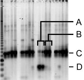
Figure 2. Dilution series of non-deleted vs. deleted DNA. The poison primer method allows detection of the deleted PCR product even in mixtures as complex as 5000 normal/1 deleted genomes. Poison Requirement for Deletion Detection in a Library Screen Figure 3 illustrates a requirement for the poison primer in detection of a 341 bp deletion (arrow) in a mutant library. The MW marker is a 100 bp ladder. The wild-type band runs at approximately 1.2 kb. This mutation was detected in a screen of a pooled 20,000 genome library, in a pool complexity of 240:1 (10 F1 worms per starting population, 1152 populations, collapsed to 96 pools for screening). The deletion band is clearly visible in lanes correspong to PCR amplifications that had the poison primer in the first-round reaction mix, but cannot be observed in lanes corresponding to those that lacked the additional primer. ? Figure 3. The poison primer method enhances detection of deletion PCR products. Poison (A) and standard (B) screening PCRs were performed in a complex population of wild type and deleted DNA. Band C corresponds to the wild type (undeleted) PCR product. Band D is the deletion PCR product. Note that the deletion product was not detected with the standard screening reaction. Summary One must recognize that this strategy changes the target for deletion relative to our typical strategy. Using our original protocols, we can detect deletions larger than 600 bp in an interval of 3000 bp. With the alternative poison primer method the deletion must eliminate the internal poison primer site. In a 3000 bp interval, therefore, one can detect only about 7% of the total number of 100 bp deletions, 14% of the 200 bp deletions, etc. In practice therefore, similar numbers of PCR reactions are required in the two approaches to recover a deletion allele. The poison primer approach, however, gives greater control over the position of the deletion within the target. This will become increasingly critical as researchers desire not just a deletion in a region but rather a precise designer deletion. For example, eventually researchers will require deletions that eliminate only a single exon in an alternatively spliced gene, or the removal of a small part of a promoter, or even the elimination of a gene that lies within the intron of another gene. This new method should make these possibilities an achievable goal. Acknowledgements We thank Marco Marra and Steven Jones of the Genome Sequence Center in Vancouver BC for their advice and enthusiasm for this project, and for the use of their Fluorimager to scan gels. This work was supported by a National Institutes of Health grant RO1HG01843 to R.B., and a grant from the Canadian Institute for Health Research to D.M. |
→如果您认为本词条还有待完善,请 编辑词条
上一篇Mitotic chromosomes from mouse peripheral blood 下一篇人类的皮肤纹理分析(图)
词条内容仅供参考,如果您需要解决具体问题
(尤其在法律、医学等领域),建议您咨询相关领域专业人士。
0
收藏到: 
 ?
? ?
?