|
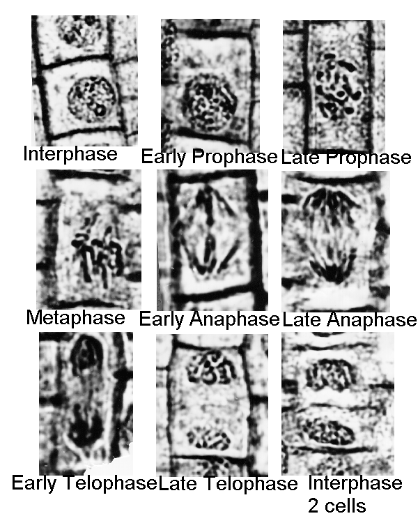
Materials Prepared slide of Onion root tip Prepared slide of whitefish blastula Microscope Procedure 1.Obtain a slide of an onion root tip and examine it for the basic stages of mitosis. Refer to Figure 11.3 for comparison. 2.Draw and label each of the stages. 3.Obtain a slide of a whitefish blastula for observation of the stages of mitosis in an animal cell. Since early embryogenesis involves rapid cellular division, the whitefish blastula has long served as a model of mitotic division in animals. It also has the advantage of demonstrating clear spindle formation in the cytoplasm. 4.Using Figure 11.3A as a guide, draw and label all stages of mitosis, including spindle formation, in the whitefish blastula. 5.Compare the division of the cell (cytokinesis) from the onion root tip to that of the whitefish blastomeres. |
→如果您认为本词条还有待完善,请 编辑词条
上一篇细胞培养从头学 下一篇视神经少突胶质细胞体外培养纯化及鉴定
词条内容仅供参考,如果您需要解决具体问题
(尤其在法律、医学等领域),建议您咨询相关领域专业人士。
0
收藏到: