|
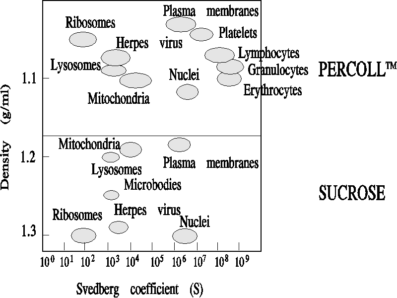
Materials Ultracentrifuge and swinging bucket rotor Procedure 1.For your ultracentrifuge, obtain a series of centrifuge tubes containing appropriate volumes of Percoll starting at 1.08 g/ml density as follows: 2.Use a hemacytometer to calculate the number of cells per ml of your blood sample. Carefully layer a suspension containing about 100 x 10 blood cells onto the gradients in tubes b-d. 3.Remove the tubes, fractionate into 0.5 ml fractions and count the number of cells in each fraction with a hemocytometer. 4.Graph the distance from the bottom of the tube vs. the number of cells in each fraction. 5.Overlay this graph with a comparison of distance vs. density of the medium, as determined by the position of the colored beads in your control tube. 6.Compare your results to Figure 1. Notes Pertoft and coworkers developed a synthetic, colloidal solution of polyvinylpyrrolidone coated silica, specifically designed for sedimentation centrifugation. This material is marketed under the trade name of Percoll. Table 1 give the characteristics of this medium, compared to several other density media, namely sucrose, metrizamide and FicollTM. Of particular interest is the fact that during centrifugation in a fixed angle rotor, PercollTM will spontaneously form linear gradients, the shape of which is dependent upon rotor speed and time of centrifugation. Thus, it becomes a simple matter to establish a linear density gradient. Figure 1 demonstrates a comparison of Percoll TMfractionation of cellular components to sucrose fractionation. This figure also presents, a standard way to comare components, by defining the relationship of density to the sedimentation coefficient for a cell or organelle. Note, that lymphocytes, granulocytes and erythrocytes have very similar sedimentation coefficients, but can be separated on the basis of density. Organelle separations are much easier to accomplish on Percoll density gradients than on sucrose gradients. Figure 1 Svedberg Values in Percoll and Sucrose
Table 1 Densities of cell structures in sucrose
|
词条内容仅供参考,如果您需要解决具体问题
(尤其在法律、医学等领域),建议您咨询相关领域专业人士。
0
收藏到: