|
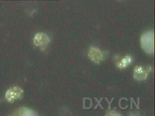
这幅图是网上的,我想找到他的染色方法,可惜始终没找到。我给出来主要是给各位看荧光显微镜下凋亡小体的样子,这个问题曾经困惑我很久,在生命经纬也求助过,未果!战友们有兴趣可以看看。 http://www.biox.cn/bbs/post/view?bid=66&id=1109620&tpg=1&ppg=1&sty=1&age=0#11096 生命经纬网友IRAM认为: 我想这副图片的细胞染色法应该是AO-EB染色法吧。因为我在做这种方法,觉得很简单,但效果明显,图片漂亮,只是假阳性稍高。 Promega公司的DeadEnd™比色法细胞凋亡检测系统能标记片断化的DNA,而这种片段化的DNA被视为是细胞凋亡的典型生化特征之一。DeadEnd™比色法细胞凋亡检测系统是一种在组织切片与培养细胞中标记凋亡细胞核的理想方法,与此同时,还可以做形态学上的评估。本文中,将展示此种方法在细胞凋亡的原位细胞, 动物模型及多种病理学组织切片中的应用。 原文:http://www.promega.com.cn/notes/emgzf/2003-4/5.htm 抗Fas单克隆抗体(50ng/ml;克隆CH-11,Oncor)诱导的Jurkat细胞的细胞凋亡。DeadEnd™比色法细胞凋亡检测系统标记Fas单克隆抗体处理过的细胞核(16小时),而未处理过的则未被染色。 |
|
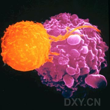
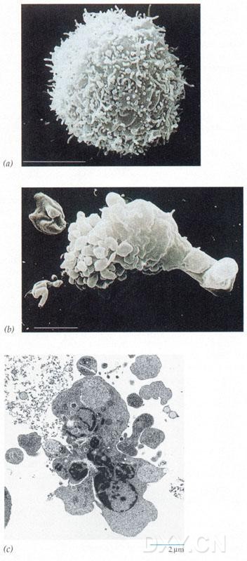
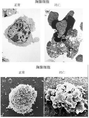
Apoptosis is of Greek origin meaning "falling off". The term is used in an analogy to the apparent suicide of leaves resulting in the very visible colour changes associated with the Autumn/Fall and that eventually leads to the leaves falling from the trees. Similiarly, cells go through a preordained sequence of events resulting in death and removal from the body. Apoptosis is a regimented sequence of events, with each act being executed in a timed fashion. Its orderliness is often referred to as the "dance of death". Microscopic examination of the cell reveals that there are events occurring both externally and internally that can be listed as follows: Externally: Shrinkage or loss of volume of the cell Blebbing on the surface of the cell Externalisation of a phospholipids termed phosphatidylserine Internally: Condensation of cytoplasm Breakdown in mitochondria integrity with the subsequent release of apoptosis inducing factors such as cytochrome c Activation of caspases Degradation of chromosomal DNA into 180-bp internuclosomal fragments Degradation of a large number of proteins (>100) thought to be important in the cell survival and metabolism 这应该是个免疫组化的片子吧,我没做过 Two human tumour cells, one normal (left) and the second (right) undergoing apoptosis following exposure to cytotoxic drugs DeadEnd™比色法细胞凋亡检测系统实验流程 |
|
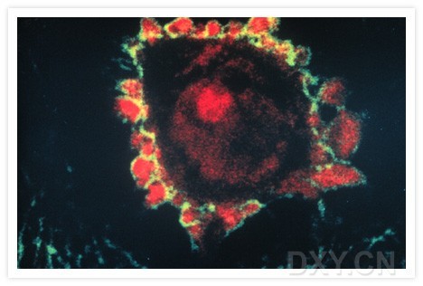
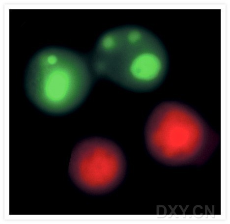
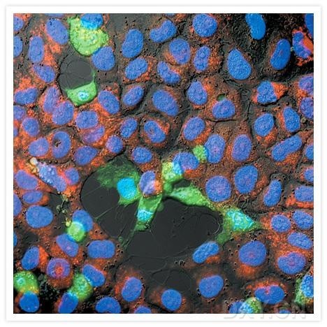
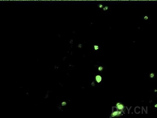
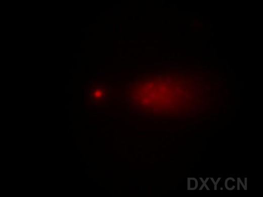
What happens during apoptosis? To kill itself, a cell first makes a deadly chemical cocktail. It then separates itself from its neighbours, and unleashes the poisons. These include a substance that chews up the DNA in the cell nucleus, and a 'glue' that binds the inside of the cell together. Within a few hours, the cell shrinks, breaks up and is engulfed by other cells. A cancer cell (mauve) undergoing apoptosis. Dr Andrejs Liepins/Science Photo Library Apoptotic human keratinocytes that were fixed with paraformaldehyde and stained with fluorescein concanavalin A (C827). The cells were subsequently permeabilized with acetone, stained with propidium iodide (P1304, P3566, P21493) and visualized by confocal laser-scanning microscopy. This photomicrograph clearly shows that the green-fluorescent lectin staining outlines the apoptotic surface blebs, whereas the red-fluorescent propidium iodide stains the interior of the blebs. Image contributed by Livia Casciola-Rosen and Antony Rose, Johns Hopkins University. Reproduced from by copyright permission of the Rockefeller University Press. 凋亡的人类角化细胞被多聚甲醛固定,被荧光素标记的刀豆蛋白A染色。然后经丙酮渗透处理,被pi染色后能被共聚焦显微镜观察。本片显示的绿色荧光(外源性凝集素)勾勒出凋亡细胞表面的小泡,红色荧光(pi)被包被在其中。 Apoptosis induced in Jurkat cells with 10 µM camptothecin. The cells were then treated with the reagents in the Vybrant Apoptosis Assay Kit #4 (V13243). Apoptotic cell nuclei were labeled with YO-PRO-1 dye (green) (Y3603). Necrotic cells were detected with propidium iodide (red) |
|
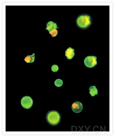
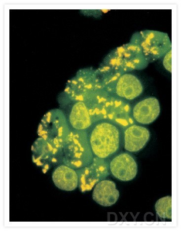
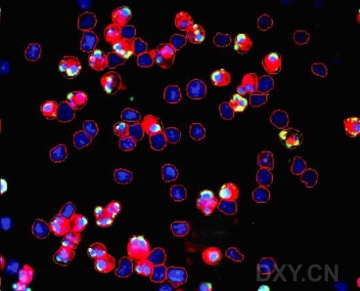
10 µM喜树碱诱导的Jurkat cells(慢淋。G1检测点缺陷细胞) 凋亡。细胞然后经 Vybrant Apoptosis Assay Kit #4 (V13243).处理。凋亡细胞核被YO-PRO-1绿染,坏死细胞被pi红染。 Jurkat human T-cell leukemia cells treated with 1 µM camptothecin. The externalized phosphatidylserine, a characteristic of early-stage apoptotic cells, was detected with Alexa Fluor 488 annexin V (A13201). The late-stage apoptotic and necrotic cells were stained with propidium iodide (P1304, P3566, P21493). The image was acquired using bandpass filters appropriate for fluorescein. Jurkat 人类白血病T细胞经1微升喜树碱处理,ps外翻,早期凋亡特异性的特征被annexin-5检测到。晚期凋亡和坏死细胞被pi染色。本图是由相应的荧光滤光片获取的。 HL-60 cells treated with camptothecin for three hours. The DNA strand nicks characteristic of apoptosis were detected with the TUNEL (terminal deoxynucleotidyl transferase–mediated dUTP nick end-labeling) assay using the fluorescently labeled nucleotide, ChromaTide BODIPY FL-14-dUTP (C7614). Image contributed by Zbigniew Darzynkiewicz, Cancer Research Institute, New York Medical College. |
|
HL-60 cells(急粒,p53缺陷)被喜树碱诱导3小时。DNA段端被荧光标记的脱氧核苷酸末端转移酶标记。 Detection of apoptosis in SK-N-MC neuroblastoma cells. Following a six-hour exposure to hydrogen peroxide, cells were labeled with Hoechst 33342 (H1399, H3570, H21492), tetramethylrhodamine ethyl ester (TMRE, T669) and rhodamine 110, bis-L-aspartic acid amide (R22122) for 15 minutes. Apoptotic cells show green cytosolic fluorescence resulting from cleavage of the rhodamine 110, bis-L-aspartic acid amide substrate by active caspase-3. The staining pattern of the Hoechst 33342 dye reveals that the majority of the rhodamine 110–positive cells also contain condensed or fragmented nuclei characteristic of apoptosis. Furthermore, the rhodamine 110–positive cells are also characterized by an absence of polarized mitochondria, as indicated by their failure to load the positively charged mitochondrial indicator TMRE. The image was contributed by A.K. Stout and J.T. Greenamyre, Emory University. 线虫发育中细胞的凋亡: 用共聚焦显微镜观察线虫胚胎,在正常胚胎CED-4位于线粒体。而凋亡细胞CED-4(红色)和核纤层蛋白(绿色)在核被膜(黄色)均可见。该实验有助于揭示,在细胞凋亡中CED-4从线粒体易位到核被膜 |
|
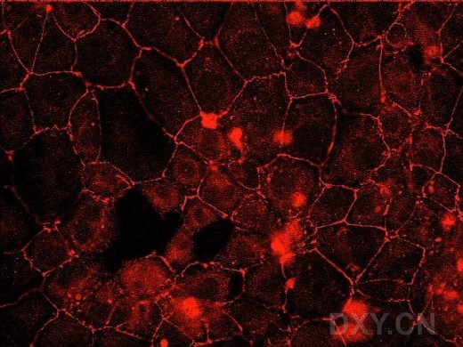
A comparison of normal and apoptotic cells. Double label ZO-1 immunofluorescence and apoptosis staining (tissue culture cells). Rinse coverslips 1X in PBS. Fix in -20oC methanol for 20 minutes Keep Coplin jars in ethanol-dry ice bath. Wash 3X 5 minutes in PBS. Block with 10% goat serum in PBS for 30 minutes at RT. Incubate with primary antibody. Rabbit polyclonal anti-ZO-1 from ZYMED, 1:200 in PBS + 10% goat serum. Incubate for 1hr at 37oC in humid chamber. Wash 3X 20 minutes in PBS. Postfix in 4% paraformaldehyde for 10 minutes at RT. Use 16% methanol free paraformaldehyde from Polysciences, diluted 1:4 in 1XPBS. Best done by using 4ml 10X PBS + 1 ampoule of 16% paraformaldehyde (10ml) and adjust the volume to 40ml by DI water. Wash in PBS 1X5 min. Block aldehydes in 100mM glycine in 0.5X PBS (pH 8.0) for 30 min. at RT. Wash in PBS 2X5 min. Block with 10% goat serum in PBS for 30 minutes at RT. Incubate with secondary antibody. Anti-rabbit IgG-Texas red conjugate (ICN), 1:100 in PBS + 10% goat serum. Incubate for 1hr at 37oC in humid chamber. Incubate in 0.2% Triton X-100 in PBS for 5 min. at RT. Wash 3X 20 minutes in PBS. Remove excess liquid by tapping the slides and touching the edge of slides to absorbent paper. Cover cells with 100 µl equilibration buffer and a piece of parafilm, to prevent drying and to spread the buffer evenly. Incubate at RT 5-10 min. Remove parafilm and equilibration buffer from slides and touch the edge of slides to absorbent paper. Incubate slides with labeling mix at 37oC for 60 min. covered with parafilm. Labeling mix for a single reaction: 45 µl equilibration buffer, 5 µl nucleotide mix and 1 µl TdT enzyme. Multiply volumes with the number of samples. Wash slides in 2XSSC for 15 min. at RT. Immerse slides in Hoeshct solution for 10 min. at RT (1:5000 dilution of 1mg/ml stock in PBS). Wash slides 3 X 5 min. in PBS. Mount slides using anti-fade from Molecular probes. If your procedure was successful, your result should look something like this |
|
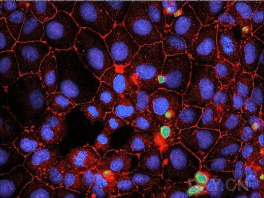
IEC-6 rat small intestinal epithelial cells were grown on glass coverslips and processed as described above. The three individual images in the following represent pseudo-colored fluorescence micrographs of nuclear stain with Hoeshct(top ), apoptosis stain with TUNEL staining (middle) and ZO-1 indirect immunofluorescence (bottom). The last image is a digital overlay of the three individual images. Staining was prepared by Luba Adler, MS. middle bottom last one 来源:http://pubweb.nwu.edu/~tji796/users/jilling/zo1apo.htm |
|
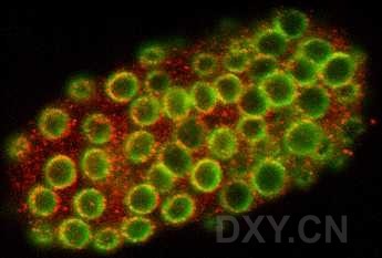
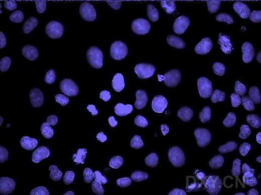
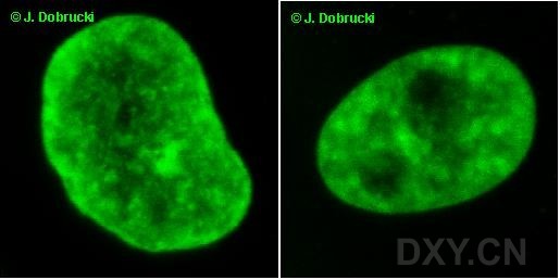
a microscope image of a group of normal cells and one apototic cell which binds annexin (green) on the surface. Annexin is labeled with fluorescein. In apoptosis condensation and fragmentation of chromatin occurs. Subsequently, nuclei loose their round or oval shape, bud and become fragmented - apoptotic bodies are formed Nuclei of normal human fibroblasts in culture, in early spontaneous apoptosis. Nuclei of normal human fibroblasts in culture, in advanced spontaneous apoptosis |
|
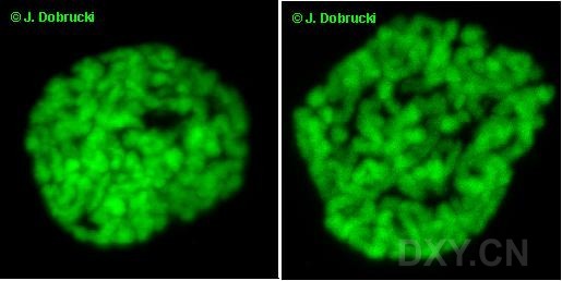
Apoptosis caused in HL60 cells by camptothecin (topoisomerase inhibitor). Images of nuclei in early apoptosis. Apoptosis caused in HL60 cells by camptothecin (topoisomerase inhibitor). Images of nuclei in mid apoptosis Apoptosis caused in HL60 cells by camptothecin (topoisomerase inhibitor). Images of nuclei in late apoptosis |
|
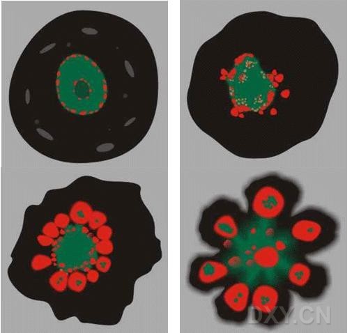
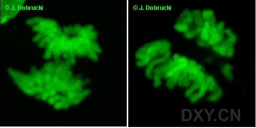
A schematic view of a nucleus in a cell undergoing apoptosis: healthy cell (top, right), early apoptosis (top, left), advanced apoptosis (bottom, right), late apoptosis (bottom, left). Subsequent stages of the cell division cycle. Histone H2B was tagged with green fluorescent protein (GFP). This made it possible to obtain fluorescence confocal microscopy images of chromatin. Interphase nuclei Prophase nuclei |
|
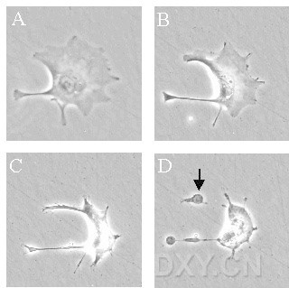
Anaphase nuclei Figure , which shows time-lapse microscopy images of a trophoblast cell undergoing apoptosis 海马CA2-3区神经细胞原位凋亡检测(TUNEL法,400X) |
|
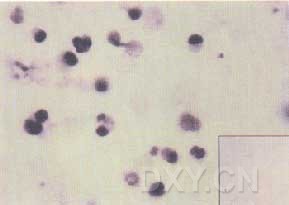
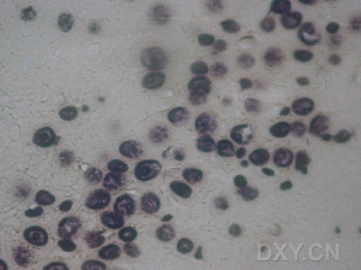
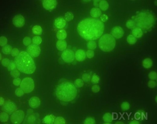
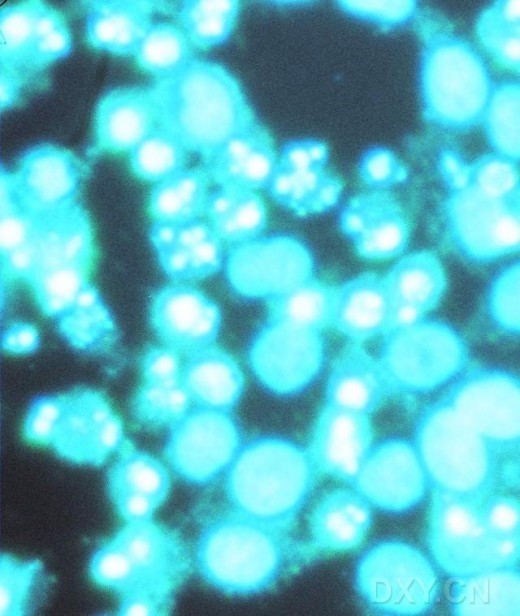
原位细胞凋亡检测(TUNEL)法: 1、石蜡切片常规脱蜡至水。 2、切片滴加0.1%TritonX-100于4℃孵育5分钟,PBS缓冲液洗5分钟×3次。 3、每张切片滴加TUNEL标记反应混合液50ul,置于湿盒中,37℃孵育1小时。 4、PBS缓冲液洗5分钟×3次,擦干组织切片后,每张切片滴加AP转化液50ul,置于湿盒中,37℃孵育30分钟。 5、PBS缓冲液洗5分钟×3次。 6、每张切片滴加BCIP/NBT显色液15-50ul,室温避光显色10-30分钟,光镜下观察。 7、镜下显色满意后,PBS缓冲液洗2-3次终止显色反应。梯度酒精脱水、二甲苯透明后,中性树胶封片。 8、光镜下观察,凋亡阳性细胞为新月型或蚕豆状浓染,并可见蓝紫色凋亡小体。 (NBT : 氮蓝四唑 BCIP: 5-溴-4-氯-3-吲哚-磷酸二钠) apoptosis hoechst33258染色的K562细胞.(凋亡) 用彗星试验观察细胞凋亡。 形态学:彗星呈明显的小头大尾。 原理:凋亡细胞产生的DNA片断大小相对均匀,所以出现在尾部的某一部分DNA片断较多,彗星尾较宽。此类图像在专题总结子版已经帖过了,这里为对比再帖一下,并附上一张非凋亡细胞的彗星图像的链接,(对照观察更直观)。 |
|
调亡细胞 周围有花瓣样结构的细胞正在调亡 |
|
凋亡细胞,用HOECHST33342染色,在荧光显微镜下观察 Human lymphoma cells treated with camptothecin for four hours and stained using the APO-BrdU TUNEL Assay Kit (A23210). Cells containing DNA strand nicks characteristic of apoptosis are detected by TUNEL and fluoresce green, while necrotic cells are stained with red-fluorescent propidium iodide. |
→如果您认为本词条还有待完善,请 编辑词条
词条内容仅供参考,如果您需要解决具体问题
(尤其在法律、医学等领域),建议您咨询相关领域专业人士。
0
收藏到: