|
Introductory Concepts When specimens, living or non-living, organic or inorganic, absorb and subsequently re-radiate light, we describe the process as photoluminescence. If the light emission persists for up to a few seconds after the excitation light is withdrawn, the phenomenon is known as phosphorescence. Fluorescence, on the other hand, describes light emission which continues only during the absorption of the excitation light. The time interval between absorption of excitation light and emission of re-radiated light in fluorescence is of extraordinarily short duration, usually less than a millionth of a second.
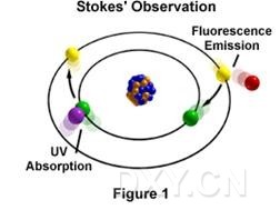
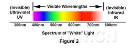
The phenomenon of fluorescence was known by the middle of the 19th century. It was Stokes who made the observation that the mineral fluorspar fluoresces when ultraviolet light is directed upon it; he coined the word "fluorescence". Stokes observed that the fluorescing light is in longer wavelengths than those of the excitation light as illustrated in Figure 1 above. In this figure, a photon of ultraviolet radiation (purple) collides with an electron in the atom, exciting and elevating it to a higher energy level. Subsequently, the excited electron relaxes to a lower level and emits light in the form of a lower-energy photon (red) in the visible light region. Figure 2 is a diagrammatic representation of the visible light region of electromagnetic radiation, which covers a wavelength range of approximately 400 to 740 nanometers. Surrounding the visible region is higher energy ultraviolet light and lower energy infrared light.
Fluorescence microscopy is an excellent method of studying material which can be made to fluoresce, either in its natural form (primary or autofluorescence) or when treated with chemicals capable of fluorescing (secondary fluorescence). The fluorescence microscope was devised in the early part of the 20th century; Koehler, Reichert, and Lehman were among the scientists associated with such development. However, the potential of this instrument was not realized for several decades. Early investigations showed that many specimens (microminerals, crystals, resins, crude drugs, butter, chlorophyll, vitamins, inorganic compounds, etc.) autofluoresce when irradiated with ultraviolet light. However, it was not until the 1930's that Haitinger and others developed the technique of secondary fluorescence--employing fluorochrome stains to stain specific tissue components, bacteria, or other pathogens which do not autofluoresce. These fluorochrome stains, tagged to specific objects, spurred the use of the fluorescence microscope. The instrument's value was significantly enhanced by the 1950's when Coons and Kaplan demonstrated the localization of antigens in tissues that were stained with a fluorescein (the fluorochrome) tagged antibody. 《Fluorescence Microscopy》下载 点击这里下载 |
→如果您认为本词条还有待完善,请 编辑词条
上一篇薄层色谱法的相关知识简介 下一篇冷冻切片机的基本操作
词条内容仅供参考,如果您需要解决具体问题
(尤其在法律、医学等领域),建议您咨询相关领域专业人士。
0
收藏到: