|
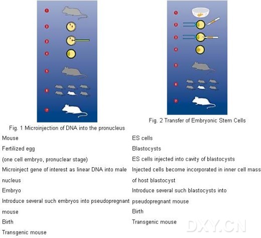
The term “transgenic animals” describes animals whose chromosomes contain stably integrated copies of genes or gene constructs derived from other species or not normally found in the host animal. For this reason, mice, rats or other small mammals are used to introduce foreign DNA into oocytes or embryos (blastocysts). Two different techniques are used: Microinjection of linear DNA into the pronucleus
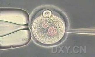
Microinjection of DNA Injection of linear DNA molecules into fertilized eggs (pronuclear stage) using an inverted microscope, micromanipulation equipment and injection / holding devices.
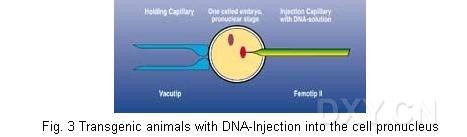
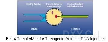
The first successful production of transgenic mice using pronuclear microinjection was reported in 1980 [1]. The pronuclear microinjection method of producing a transgenic animal results in the introduction of linear DNA sequences into the chromosomes of the fertilized eggs. If this transferred genetic material is integrated into one of the embryonic chromosomes, the animal will be born with a copy of this new information in every cell. The foreign DNA must be integrated into the genome prior to the doubling of the genetic material that precedes the first cleavage. If this does not occur, only a few cells will integrate the gene. For this reason, the DNA is introduced into the fertilized egg at the earliest stage, which is the pronuclear period immediately following fertilization. For several hours following the entry of the sperm into the oocyte, the male and the female pronuclei are visible as individuals under normal light microscope and not fused into a so-called zygote. The DNA may be injected into either of these pronuclei with no difference in results. Usually the injection is into the male pronucleus because it is larger than the female nucleus and also because it is closer to the oocyte surface. These oocytes are subsequently transferred into the uterus of pseudopregnant recipient animals and develop to term. Setup of the workstation: One TransferMan micromanipulator is used for moving the holding capillary, the other TransferMan is used for moving the injection capillary. The actual holding of the embryo is carried out using CellTram Air. Two possibilities are available for injecting DNA: using the CellTram Air as a simple injection device or using the FemtoJet™ for reproducible and programmable injections. Together with an inverted microscope, combined with Hoffmann modulation contrast optics or differential interference contrast optics, microinjection can be effected quickly and simply. Preparation of the object slide: One drop of medium is placed on the slide. The embryos to be injected (10 to 20) are placed in one area. The whole droplet is covered with oil. Operating: Operating TransferMan, by means of four self-explanatory keys, is extremely simple. For every TransferMan device, only two spatial coordinates are defined and stored. The capillary can be moved easily in any direction (x y z) by means of a joystick. By simply pressing the joystick button, the capillary can be moved repeatedly to one of the two pre-programmed positions. The setting of these positions depends on how the TransferMan is used (Fig. 4)
|
|
Microinjection is carried out as follows: An embryo is brought into the injection position using the holding capillary. The tip of the injection capillary is aligned with the pronucleus and inserted directly into the pronucleus. Approximately 1 pl DNA solution is injected during every injection process. The injection can be considered successful when the pronucleus increases in size. If the volume of the pronucleus does not increase, the injection is considered unsuccessful. The injection would then occur into cytoplasm, where the injection drop shows different contrast. If the tip comes too close to the endogenous DNA during the injection process, the DNA sticks to the capillary when it is pulled out. If this occurs, the capillary should be discarded. The correct injection volume of the Transjector is set by defining the injection time and the injection volume for all embryos. Femtotips and Microloaders Femtotips, the standard capillaries for microinjection, are ideal for the injection of DNA, RNA, proteins and dyes into living cells. The shapes of the capillary tip ensures high cell viability and minimizes blockages by the injection material. The capillary opening is constant, allowing reproducible injection volumes in the femtoliter range. Femtotips are available in two shapes for injecting cultured cells or suspended cells. Backfilling with an Eppendorf Microloader easily places the injection material directly into the end of the Femtotip. Transfer of Embryonic Stem Cells - Injection of ES cells into the cavity of blastocysts using a microscope (inverted), micromanipulator equipment and transfer / holding devices [2]. Embryonic stem cells are derived from the inner cell masses of normal blastocysts (early embryos, mostly mouse embryos). These cells are pluripotent, which means they can develop into any type of tissue. Therefore, removing ES cells from the culture and placing them back into an early embryo (blastocyst) allows them to divide and become part of the embryo. Transfection allows foreign genetic material to be inserted in vitro into these cells. The target of this process is homologous recombination with the chromosome of the cell, i.e. introduction or exchange of DNA at one location with homologous DNA sequences. Following transfection, the descendants of one individual cell (clone) are analyzed using microbiological methods to ascertain whether any mutation is present. Cells of the identified clones which showed the desired reaction are multiplied in vitro and injected into blastocysts. These injected blastocysts are then implanted into the uterus of pseudo-pregnant females. Setup of the workstation: As for the injection of DNA, two TransferMan devices are used for controlling the holding and transfer capillaries. The actual holding of the blastocysts is performed by using CellTram Air. CellTram Oil is used for transferring the ES cells. As aforementioned, one inverted microscope, in combination with Hoffmann modulation contrast optics or differential interference contrast optics, makes the ES cell transfer quick and simple. The setting of the positions is carried out as shown in Fig. 6: Right-hand side - injection position: The area where the transfer capillary is kept for injection (focus level, magnification x20 or x40 times) is defined as Position 1. Position 2 is its “parking” position. This can be outside and above the droplet (in the overlay medium). This capillary holder is connected to CellTram Oil. Left-hand side - holding function: The area where the uninjected blastocysts are placed is set to Position 1. Here one blastocyst can be easily taken by using CellTram Air and VacuTip. This blastocyst is moved via the joystick to a central position to inject the ES cells. After ES cells have been injected, the capillary is moved to a “parking” position (can be outside the view). This position is defined as Position 2. Pressing the joystick button moves the capillary back to Position 1. The procedure then restarts. Preparation of the petri dish: Blastocysts are placed onto a petri dish in one or more drops of medium. Blastocysts should be positioned close together in one area of the drop. The ES cells are placed in another drop. All drops on the dish are covered with oil. The method of procedure is as follows: The transfer capillary is directed to the focus level and the position is stored as Position 1. Position 2 is defined beside or above this drop. The drop with the ES cells is placed in focus and several ES cells are aspirated into the transfer capillary by rotating the knob of the CellTram Oil. The transfer capillary, which now contains the cells, is moved up into the overlay medium. The petri dish is moved in order to bring a blastocyst in the other drop into view. One blastocyst is taken with the second TransferMan (the holding capillary) in the Position 1 and moved to the injection position. In this position, the ES cells are injected into the cavity of this blastocyst. The injected blastocyst is stored in Position 2 by simply pressing the joystick button of the manipulator on the left-hand side. The automation of repetitive tasks while maintaining easy operation allows the user to concentrate on the micromanipulations themselves. This enables rapid work with good results. Replace one TransferMan with an InjectMan and expand the versatility of the system to include adherent cell injection. Literature [1] Gordon, J.W., Scangos, G.A., Plotkin, D.J., Barbosa, J.A., Ruddle, F.H.: Genetic transformation of mouse embryo by microinjection of purified DNA. (1980)Proc.Natl.Acad.Sci USA 77, 7380-7384 |
→如果您认为本词条还有待完善,请 编辑词条
词条内容仅供参考,如果您需要解决具体问题
(尤其在法律、医学等领域),建议您咨询相关领域专业人士。
0
收藏到: