|
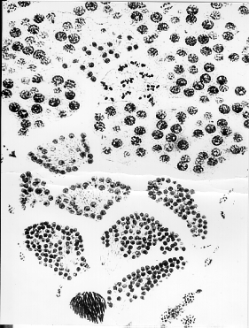
Materials Prepared longitudinal section of grasshopper testis Microscope Procedure 1.Place the slide on the microscope and with low power identify the apical end of the testis and the region where the testis joins with the vas deferens. The apical end is round and packed with cells, while the opposite end is a more open cavity lined with formed sperm. 2.Identify the individual compartments of the testis, known as cysts. The cysts are separated by connective tissue walls or septa. 3.At the apical end, the cells are grouped into presumptive germ cells known as spermatogonia. These cells are undergoing mitosis and are giving rise to all of the remaining germ cells in the testis. Moving from the spermatogonia in the apical end to the vas deferens at the opposite end of the testis, the cells mature as a group. That is, all of the cells within a given cyst will be in the same approximate stage of meiosis. A good longitutinal section will have nearly all of the stages of meiosis displayed on one section, but you may have to examine several slides to observe all of the stages. 4.Using Figure 11.6 as a guide, identify , draw and label each of the following stages: Spermatogonia Primary spermatocytes in the following phases of Prophase I leptotene zygotene pachytene diplotene diakinesis. Secondary spermatocytes Spermatids Sperm |
→如果您认为本词条还有待完善,请 编辑词条
上一篇我养lovo细胞的经验 下一篇酵母表达系统
词条内容仅供参考,如果您需要解决具体问题
(尤其在法律、医学等领域),建议您咨询相关领域专业人士。
0
收藏到: