标签: Western-blot 蛋白杂交
顶[0] 发表评论(10) 编辑词条
|
Western blot is a powerful technique in that; it leads to simultaneously detection of a specific protein by means of its antigenicity and its molecular mass. The proteins are first separated by mass using SDS-PAGE, transferred from gels onto membranes and then specifically detected in the immunoassay step. Information on activation status (i.e. enzyme pro-form vs. active species, unless specific antibodies against the pro species are available), oligomeric arrangement and post-translational modifications can also be deduced from this technique. The efficiency of protein transfer from the SDS-PAGE gel to the support membranes can be affected by the gel thickness and the total acrylamide concentration. Transfer buffers can also affect the efficiency of protein migration through the gel. While methanol is necessary to prevent gel swelling with heating, and to keep protein adsorbed to membrane, methanol-containing buffers often result in the decrease in pore size and may even precipitate proteins within the gel. Poor transfer can be remedied by using methanol-free transfer buffer. The choice of support membranes for protein transfer is usually determined primarily by the subsequent investigation steps to be performed. For conventional probing with antibodies, nitrocellulose (high binding capacity ~80-100 μg/cm2) or nylon (binding capacity ~480 μg/cm2) are suitable. Although nylon has an attractive binding capacity, blocking steps are usually performed at high temperatures. This may denature sensitive antigens and these membranes do not stain well with the anionic dyes required to check protein transfer efficiency. In western blot, the transfer of proteins in membranes is mostly followed by immunoassay step where specific antibody is used to probe for specific antigens. Immunoassay involves the following steps: Blocking; where the transfer membranes are blocked with a concentrated protein solution (e.g. ovalbumin, BSA, haemoglobin, non-fat milk powder) to prevent further non-specific binding of proteins [non-protein (polyvinylpyrrolidone) in the presence or absence of non-ionic detergent (such as Tween 20), may also be used for blocking]. This step is followed by incubation of the membrane in a diluted antibody solution, washing of the membrane, incubation in diluted conjugated probe antibody or other detecting reagent, further washing, and finally detection. 1. Transfer of protein to the membrane Apparatus and equipment ? Transfer cassette: Hoefer®TE series Transphor Electrophoresis Unit ? HybondTM -C nitrocellulose paper (0.45 μm pore size) supplied as 33 x 300 cm rolls ? Filter paper: Whatman 3mm or preferably blotting paper. Both nitrocellulose paper and filter paper are cut to the same size as gel using clean Blade and gloves. ? Blotting Electrodes are carbon slabs, 1 cm thick x 20 cm long x 15 cm wide, with heavy insulated leads, or patent blotting apparatus. ? Powerpack: Normally a high-amperage but low-voltage apparatus is preferred; one that is capable of supplying 500 mA – 1 A current at voltages as low as 5 V. Paul Meyers electroblotting apparatus is suitable. |
|
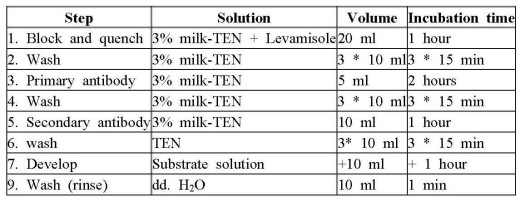
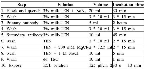
Reagents ?Gershoni blotting buffer [25 mM Tris-HCl, 192 mM glycine, 20% (v/v) methanol, 0.01% (w/v) SDS, pH 8.3]. 6.05 Tris, 28.8 g and 2 ml of 10% (w/v) SDS solution are dissolved in 1.6 litres dd.H2O. 400 ml Methanol is added and the solution is stored at -20°C without pH adjustment. ? Ponceau S proteins stain solution [0.1% (w/v) in 1% (v/v) acetic acid]. 0.1 g Ponceau S and 1 ml acetic acid are added to a 100 ml volumetric flask and made up to volume with dd.H2O. ? Tris-EDTA-NaCl buffer [TEN: 25 mM Tris-HCl, 1 mM EDTA, 150 mM NaCl, pH 7.6] 3.03 g Tris, 0.29 g EDTA and 8.77 g NaCl are dissolved in dd.H2O made up to 1 litre without pH adjustment and autoclaved. Procedure for Protein transfer 1. Following SDS-PAGE, the gel is dismantled from the electrophoresis unit, submerged briefly in ice cold transfer buffer; it is then laid flat on pre-wetted Hybond?-C nitrocellulose hybridisation transfer membrane (0.45 μm) supported on three layers of transfer buffer-wetted filter paper resting on the anode. The gel is overlaid with three wetted filter papers (or blotting paper), and then the cathode or another layer of nitrocellulose/gel/blotting paper. Care should be taken to exclude bubbles between gel and nitrocellulose, and between nitrocellulose and paper. The assembly is placed in a plastic tray resting on anode. 2. The transfer cassette (Hoefer? TE series Transphor Electrophoresis Unit) is assembled; the nitrocellulose/gel/blotting paper assembly is placed in a plastic tray resting on anode. The ANODE (electrode on the nitrocellulose paper side of the assembly) is connected to anode connector and the gel side CATHODE to the cathode connector of an appropriate powerpack. 3. The chamber is filled with ice-cold transfer buffer and stirred with a magnetic stirrer throughout the run. 4. A current of 500 mA is passed for 20-30 min to effect transfer. Transfer is performed at a constant current of 30V over night, using Paul Meyers electroblotting apparatus. 5. After transfer, the gel and the membrane are disassembled from the cassette and the outline of the gel is marked on the membrane. 6. The membrane is rinsed with dd.H2O and then rinsed with Ponceau S to visualise the MR markers, the sample proteins and to assess the efficiency of the transfer. The positions of the lanes and the markers are marled by pricking the membrane gently with a needle point. 7. The Ponceau S was decolourised with TEN and the membrane was air-dried before probing, or alternatively stored in a desiccator at 4°C. NB: Do not touch assembly while power is on!!! 2. Immunoassay
附件列表→如果您认为本词条还有待完善,请 编辑词条 词条内容仅供参考,如果您需要解决具体问题
收藏到: |